
An excellent fit to expand the imaging center, Prodiva 1.5T
FieldStrength MRI magazine
User experiences - December 2017
High quality MR imaging combined with a simplified, guided workflow
The Prodiva 1.5T is a robust, all-digital Philips MRI scanner, designed for excellent clinical performance and ease of use. With flexible coils and the simplified Breeze workflow, Prodiva 1.5T offers an enhanced user and patient experience. Prodiva 1.5T is up and running at Radiology Schiffer in Germany and Seirei Mikatahara General Hospital in Japan, where it is already exceeding expectations.
“Of course, we wanted a scanner that is easy to handle, so that it can be a workhorse for us”

Silvia Schiffer, MD Radiologist, Director of Radiology Schiffer, a private imaging center with 30 staff members. Her clinical interests lie in neurological and musculoskeletal MRI. She recently extended the facility’s imaging capabilities and capacity by relocating to a new site.
Sandra Maass Senior MRI technologist at Radiology Schiffer in Hennigsdorf, near Berlin, Germany.
Mamoru Takahashi, MD Radiologist, joined the Department of Radiology of Seirei-Mikatahara General Hospital, Japan, in 2000. His interests span the whole range of radiological image diagnosis but in recent years, he has become especially interested in MRI of the abdominal region and, in particular, of the great vessels.
Radiology Schiffer, Germany
The right choice to expand the imaging center
In the first quarter of 2017, Radiology Schiffer in Hennigsdorf, near Berlin, relocated to a new site where MRI, CT and x-ray are provided. As part of this move, the imaging center’s Director and Senior Radiologist Silvia Schiffer decided to add a second MRI system, rather than extending operation hours into evenings and weekends to meet increased capacity demands for MRI. After evaluating several options, she chose Philips Prodiva 1.5T CX as an excellent fit for her needs. Decisive factors for this choice were Prodiva’s clinical capabilities paired with the favorable price point. A compact footprint, low ceiling height and lightweight magnet meant low siting requirements and Mrs. Schiffer was also attracted by the system’s workflow enhancements and efficiency. “Of course, we wanted a scanner that is easy to handle, so that it can be a workhorse for us. And it was particularly important to me that the image quality is not inferior to what our Ingenia MRI scanner produces, because I wouldn’t want referring physicians to notice differences in quality from both systems; that would have been bad.”
"Within three days, our technologists were confidently using the new system"
Clinical cases from Radiology Schiffer
")
")
“The flexible, lightweight coils fit really well to the patient’s body shape, and the connectors are superb”
Positioning and operation: smooth and simple Once the scanner was in place, Mrs. Schiffer and her staff received tailored training in line with their clinical and workflow requirements and objectives. “I have been impressed from the start. Installation and set-up were smooth and simple. Within three days, our technologists were confidently using the new system,” recalls Mrs. Schiffer. Since installing Prodiva 1.5T, she and her team have noticed a number of enhancements to their daily work. The scanner’s simple Breeze workflow supports easy patient positioning with fewer positioning steps, rapid set-up and changeover times. Ultra-light anterior coils, short cables and small connectors are making technologists’ lives easier. Senior MRI technologist Sandra Maass says: “Speaking for myself and my colleagues, we all very much enjoy using Prodiva – and we benefit from its many workflow advantages every day.” And Mrs. Schiffer adds, “The flexible, lightweight coils fit really well to the patient’s body shape, and the connectors are superb: connecting and disconnecting requires just little effort.” “The shoulder coil is a great example of easy positioning with Prodiva,” says Mrs. Maass “Other shoulder coils can be quite rigid, so that patients with pain, or bigger stronger shoulders, or a somewhat abnormal shoulder or spine anatomy cannot be positioned well in the coil, which often leads to loss of image quality. However, the Prodiva shoulder coil is very flexible and has large coverage, which makes good positioning easier and that contributes to the superb image quality and high SNR that we get in our shoulder exams.” High quality of images impresses From a clinical perspective, too, Mrs. Schiffer is very impressed with Prodiva 1.5T. In her practice around 50% of scans are neurological, 40% musculoskeletal and the rest made up of vascular, abdominal and miscellaneous cases. The radiology team is receiving good results across anatomies, which their referring physicians are confirming to them. “Prodiva’s high image quality and advanced features, including diffusion-weighted imaging in many organs, improve our diagnostic confidence and help decision-making,” says Mrs. Schiffer. “Prodiva has been an excellent investment and is definitely the right choice for us.” “The quality of the contrast-free MR angiography keeps impressing us,” says Mrs. Schiffer. “In addition to not having to inject contrast agent, these images are amazing, for instance of the carotids. Also our exams of shoulders and hands are really exceptional. Images of the hands, for example, cover the fingertips and include the full wrist as well, all with high signal-to-noise ratio.”
“We now even perform some exams on Prodiva that we would previously only do on a 3T”
Seirei Mikatahara General Hospital, Japan
A wealth of clinical possibilities makes Prodiva a versatile system
At Seirei Mikatahara General Hospital in Shizuoka Prefecture, Japan, Prodiva 1.5T is making impact, too. Radiologist Dr. Takahashi and his colleagues in the large radiology department operate three MRI scanners, including an Ingenia 3.0T, to serve a population that includes many elderly people. When the time came to replace an older MRI system, they chose Prodiva 1.5T – and the clinical capabilities and workflow advantages of their newest scanner have had a real impact. Dr. Takahashi says: “For a 1.5T, Prodiva delivers excellent quality, which really exceeded my expectations. We now even perform some exams on Prodiva that we would previously only do on a 3T.” High SNR, motion-free images and excellent homogeneity of the magnetic field help the team achieve excellent results. “I really appreciate the quality of fat saturation with mDIXON XD, especially in the neck area and joints of extremities, which were often problematic for us,” says Dr. Takahashi. “Since introducing the Prodiva, we have had to perform fewer rescans due to the high SNR, motion-free images, and excellent homogeneity of magnetic field.” Dr. Takahashi uses Prodiva 1.5T for a variety of examinations, including cardiac, vascular, abdominal and musculoskeletal. Furthermore, he also performs whole body imaging using T1, STIR, T2WI and diffusion sequences on the Prodiva system. “I’m impressed by the short patient preparation time and fast scan time,” he says. “Typically, diffusion imaging in body is quite challenging due to distortions. We get very good results in body diffusion with Prodiva.”
Flexible coils simplify workflow From a workflow perspective, the lightweight flexible coils are easy to handle for operators. “Our operators are very pleased with the system’s Breeze workflow. The integrated neurovascular spine coil and flexible dS MSK coil help make patient set-up simple,” says Dr. Takahashi. “They are also comfortable for the large population of elderly patients at Seirei Mikatahara Hospital. And the table height is relatively low, which helps patients with limited mobility to enter the scanner easily and comfortably. In addition, thanks to the compact siting requirements of the system, we have also been able to expand our patient waiting room and control room.”
"I’m impressed by the short patient preparation time and fast scan time"
Clinical cases from Seirei Mikatahara General Hospital
")
")
")
")
")
Prodiva is designed for high throughput with low cost of ownership

Philips Prodiva 1.5T offers easy-to-use, robust imaging coupled with a smooth, highly guided workflow. The system’s Breeze workflow uses smart automation to streamline routine tasks such as table positioning and coil selection. Breeze cuts positioning steps by up to 34%1, smoothing exams and potentially helping to increase patient volume. Flexible coils and connectors make patient set-up simple. Thin, lightweight coils fit well around different patient anatomies for fast, efficient and comfortable scanning. Prodiva 1.5T boasts a small footprint which helps keep installation and siting expenses down. Prodiva can be installed even where many MRI systems cannot.2 With low cost of ownership and many ways to extract value over the long term, Prodiva 1.5T is an excellent choice for facilities looking to make the most of their investment. This durable newcomer to the Philips MR portfolio is already providing smart ways to address today’s MR challenges. Philips Prodiva 1.5T features digital dStream technology for high quality images. Digitizing the signal in the coil captures the MRI signal in its purest form. This leads to increased SNR, which can then be used to enhance image quality or reduce scan time. Powerful clinical applications provide robust, consistent quality helping physicians make confident diagnoses and treatment decisions. With the XD functionality for fat suppression, motion correction and metal artifact reduction, Prodiva 1.5T helps deliver excellent results in a short time and paves the way for high performance.
1 Based on an internal study comparing workflow with the Achieva MR system 2 Other MR refers to 60 cm 1.5T whole body MRI systems
Clinical cases from Radiology Schiffer
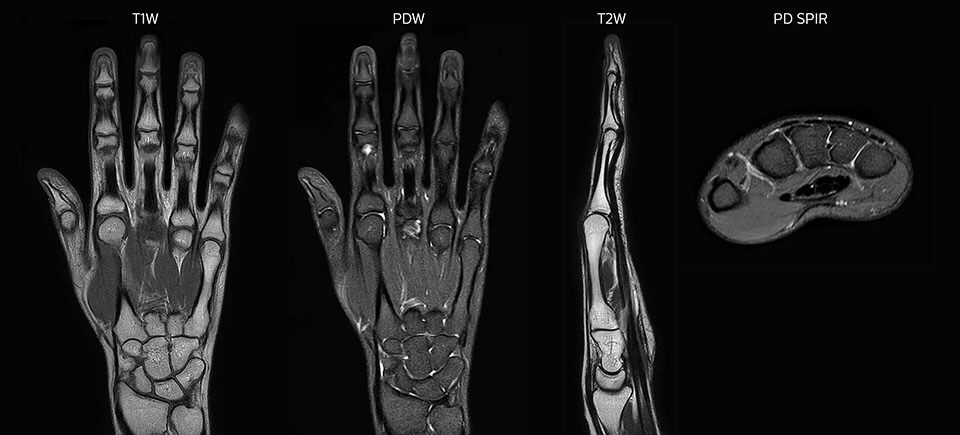
MRI of hand and wrist with large FOV
Prodiva imaging of the hand covers the fingertips and includes the full wrist as well. The dS MSK M coil is easy to use.
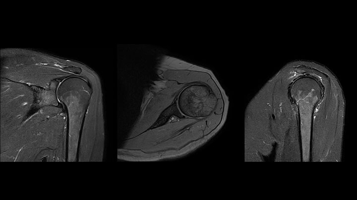
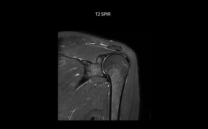
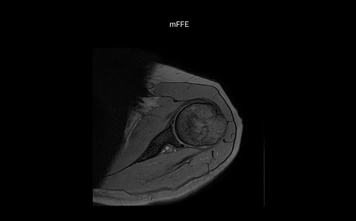
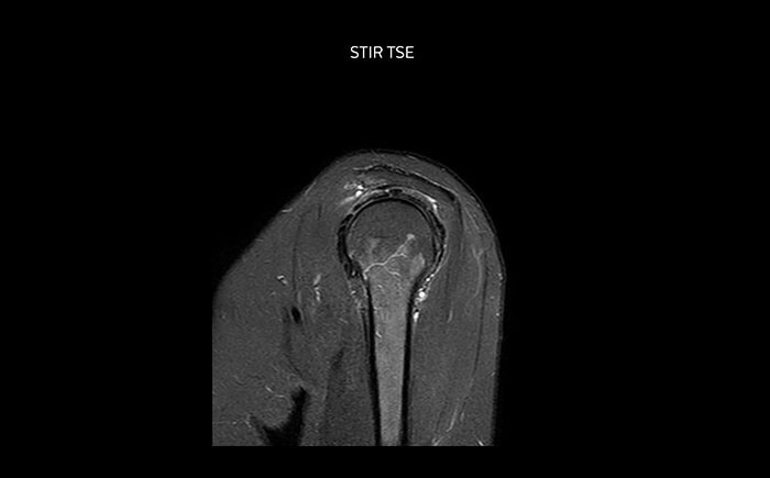
Shoulder MRI with high quality, large coverage
The Prodiva shoulder coil is very flexible and has large coverage, which makes good positioning easier, and that contributes to the superb image quality and high SNR that we get in our shoulder exams.
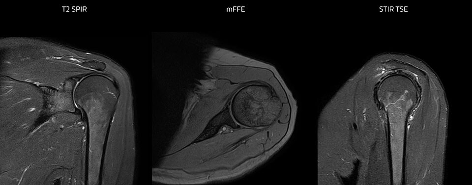
Scan time 2:55 min, FOV 160 mm, acq voxels 0.55 x 0.83 x 3.0 mm.
Scan time 4:19 min, FOV 160 mm, acq voxels 0.55 x 0.80 x 3.0 mm.
Scan time 2:50 min, FOV 160 mm, acq voxels 0.70 x 0.99 x 3.0 mm.
Shoulder MRI with high quality, large coverage
The Prodiva shoulder coil is very flexible and has large coverage, which makes good positioning easier, and that contributes to the superb image quality and high SNR that we get in our shoulder exams.
Scan time 2:55 min, FOV 160 mm, acq voxels 0.55 x 0.83 x 3.0 mm.
Scan time 4:19 min, FOV 160 mm, acq voxels 0.55 x 0.80 x 3.0 mm.
Scan time 2:50 min, FOV 160 mm, acq voxels 0.70 x 0.99 x 3.0 mm.
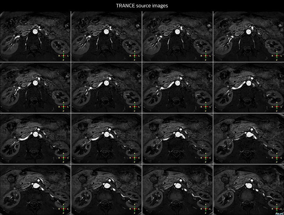
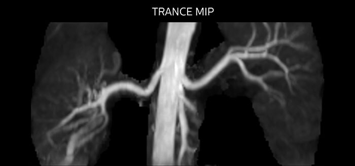
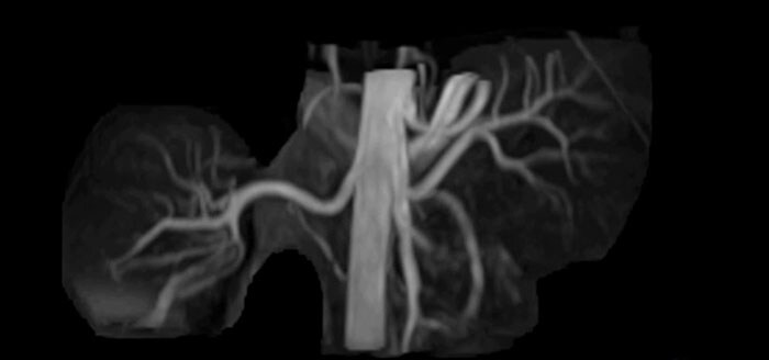
Non-contrast MRA of renal arteries
Imaging the renal arteries without contrast agent on Prodiva 1.5T.
Clinical cases from Seirei Mikatahara General Hospital
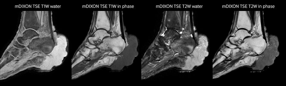
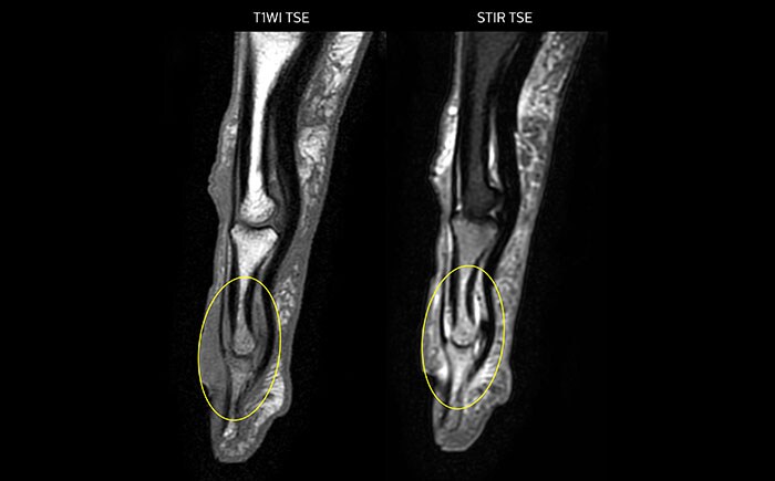
mDIXON TSE of ankle
MRI examination on Prodiva 1.5T of a 72-year-old female with a malignant melanoma in the ankle. mDIXON TSE provides excellent fat suppression, without the distortion that is often seen at such extremities.

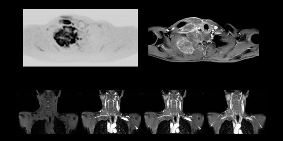
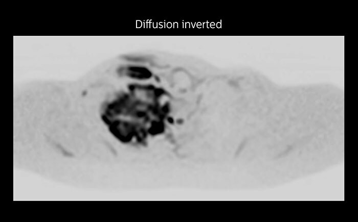
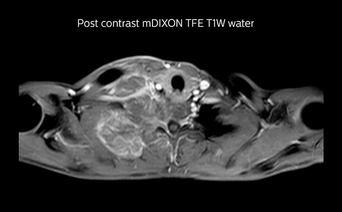
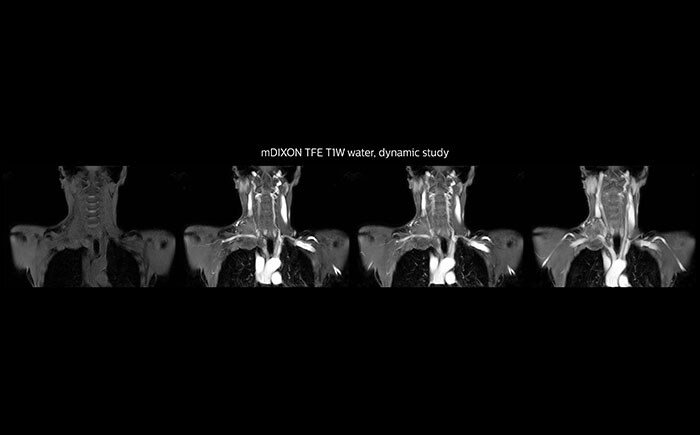
MRI of Pancoast tumor
Although the area between the neck and the top of the lung is one of the most difficult areas for MRI, Prodiva 1.5T images show good quality in this 56-year-old male with Pancoast tumor on the right. mDIXON TFE images shows excellent fat suppression in the neck area and the DWI shows almost no distortion.
MRI of the finger
MRI of the finger with high SNR and good resolution in a 10 cm field of view on Prodiva 1.5T. The diagnosis in this 63-year-old patient is bone elasmanosis.
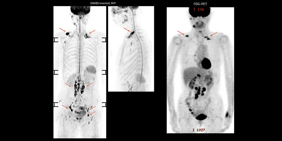
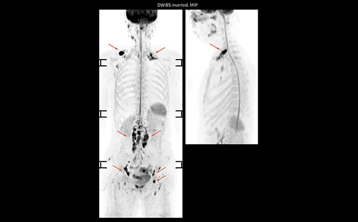
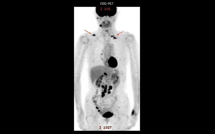
Whole body diffusion
A 61-year-old female with a malignant lymphoma underwent an MRI exam with whole body diffusion weighted imaging (DWIBS) as well as PET. On the images shown, the resolution of DWIBS is better than PET, which allows visualization of the small pelvic lesions and almost no distortion is seen in the neck area.
MR neurography of nerve sheath lesion
This patient is a 43-year-old female with a left supraclavicular nerve sheath tumor. The lesion is well visualized on the STIR VISTA images and on the MR neurography using DWIBS. The exam was performed on Prodiva 1.5T.
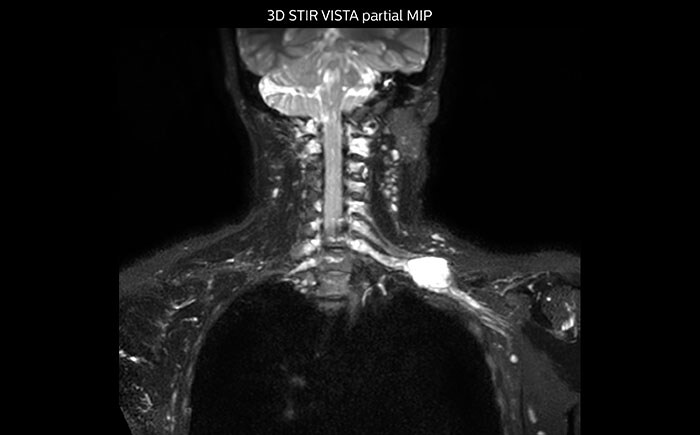
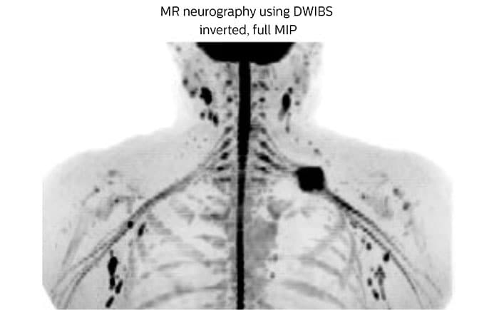
Acq voxel size 1.2 x 1.3 x 2.4 mm, Recon voxel size 0.7 x 0.7 x 1.2 mm, dS SENSE factor 2, scan time 5:46 min.
Meet
The Next MR Wave RSNA 2017
Stay in touch with Philips MRI
Related information