- Affiniti CVx: AutoStrain LV with automated EF and mid-layer strain
-
AutoStrain LV with automated EF and mid-layer strain
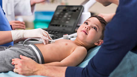
AutoStrain delivers fast, reproducible 2D strain quantification for the LV, LA, and RV, as well as automated EF and mid-layer strain for a comprehensive LV assessment within the same application, improving workflow and saving time. Smart View Select works in the background and uses AI to automatically select the optimum images for 2D LV assessment. - Affiniti CVx: 2D Auto EF and 2D Auto EF Advanced (Purchasable)
-
2D Auto EF and 2D Auto EF Advanced
Offers fast, reproducible results for easy comparison of LV functional data to improve workflow and save time in LV assessments. 2D Auto EF supports non-contrast images, and 2D Auto EF Advanced uses AI to support the quantification of contrast and non-contrast images. - Auto Segmental Wall Motion Scoring (Purchasable)
-
Auto Segmental Wall Motion Scoring
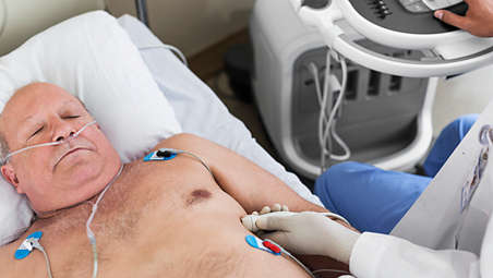
Provides automated evaluation of wall motion in a standard 17-segment bullseye display to aid objective LV wall assessment. With Auto SWMS, you can achieve greater reproducibility and efficiency in your workflows. - S5-1 PureWave Transducer
-
S5-1 PureWave Transducer
The S5-1 transducer with PureWave crystal technology generates extended operating frequency range from 5 to 1 MHz. It supports imaging in 2D, contrast mode, coronary color, CW & PW Doppler, steerable pulsed wave, High-PRF, color doppler, tissue doppler and biopsy guide capabilities. It uses reoptimized xRes adaptive image processing to provide refined tissue pattern displays, with improved details, better edges and fine border definition. It supports adult echo, abdominal, pediatric echo and TCD applications. - X5-1 xMatrix transducer (Purchasable)
-
X5-1 xMatrix transducer
Our most leading-edge, versatile ultrasound transducer technology provides remarkable 2D and 3D TTE image quality with xPlane and iRotate capabilities. Designed to simplify your imaging workflow for even difficult-to-image patients. - Dynamic HeartModel (Purchasable)
-
Dynamic HeartModel
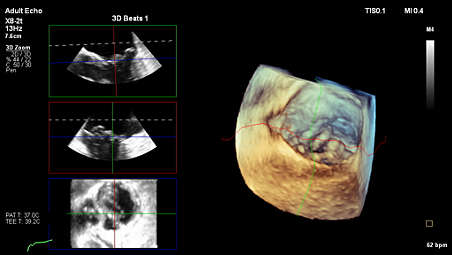
Dynamic HeartModel brings fully automated, advanced live 3D quantification. With one button press you can get 3D EF, LV and LA quantification from the same cycle . - Affiniti CVx: 3D Markers
-
3D Markers
Graphic markers can be placed within a 3D Volume or MPR while in MultiVue to streamline tracking of structural points of interest. 3D Markers facilitate greater efficiency during echo-guided procedures and allow for detailed annotation of complex anatomy. - Affiniti CVx: Auto Measure provides automation for robust, proven, reproducible cardiac quantification
-
Auto Measure provides automation for robust, proven, reproducible cardiac quantification
Auto Measure provides fully automated 2D doppler and length measurements along with AI assisted Smart (Doppler) View ID which further enhances time-savings. AI provides full functionality without EKG. - iRotate to easily access an optimal view
-
iRotate to easily access an optimal view
Electronically access the optimum view within the acoustical window between ribs instead of manually rotating the transducer to search for an unobscured window. This can increase accuracy in measuring LV volumes because the image is less likely to be foreshortened. - Next-generation TEE imaging (Purchasable)
-
Next-generation TEE imaging
The xMatrix X8-2t transducer brings exceptional image quality and confidence to TEE imaging. Live 3D and Live 3D color flow, together with latest capabilities such as xPlane Doppler and MultiVue, help you to make a confident diagnosis in even the most complex cases. - Collaboration Live for tele-ultrasound (Purchasable)
-
Collaboration Live for tele-ultrasound
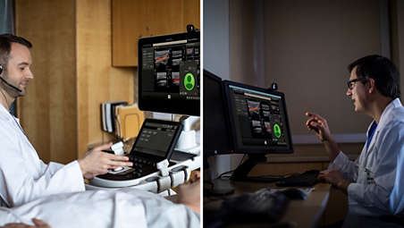
Now your ultrasound system can do more than scan. Collaboration Live allows you to reach out directly from the ultrasound system for real-time access to remote senior expertise. Collaboration Live with multi-party lets you connect up to six participants in a call. You can even connect system to system so you can give and get support from your colleagues during an ultrasound exam. - Experience echo workflow with Ultrasound Workspace
-
Experience echo workflow with Ultrasound Workspace
Ultrasound Workspace is a holistic, scalable, cardiovascular viewing, analysis and reporting system which is built on the foundation of the TOMTEC-ARENA platform. It allows for top-notch clinical efficiency by providing care teams with workflow flexibility: Enabling the same diagnostic capabilities on- and off-cart; analyzing vendor-agnostic data; leveraging AI across a wide range of applications; with a highly scalable technology platform and licensing model; and tailored comprehensive support. - Exceptional advances for pediatric imaging (Purchasable)
-
Exceptional advances for pediatric imaging
From fetal echo to pediatrics to adult congenital patients, Affiniti CVx offers a depth of imaging capability combined with streamlined cardiac workflow to reduce the steps and time needed for challenging exams such as TTE and TEE. - Elevated vascular assessment (Purchasable)
-
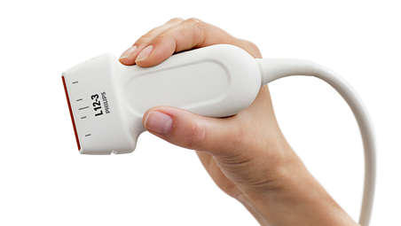
Elevated vascular assessment
The lightweight L12-3 ERGO transducer provides superb detail and resolution including MicroFlow Imaging for remarkable sensitivity in assessing blood flow. The eL18-4 transducer provides thin-slice imaging for exceptional tissue uniformity from near to far depth of field across a wide range of applications and depth requirements. - 3D Auto MV for mitral valve quantification (Purchasable)
-
3D Auto MV for mitral valve quantification
Analyze the complex anatomy of the mitral valve in 3D as well as its dynamic mechanics. This is useful from the initial discovery of mitral valve disease or pathology to support device planning, and through monitoring of pre- and postoperative cases. - 3D Auto LAA for left atrial appendage sizing (Purchasable)
-
3D Auto LAA for left atrial appendage sizing
Acquire the LAA ostium size quickly and easily. Automation reduces inter- or intra-user variability, increasing confidence during procedures. - Customizable cardiology-focused interface
-
Customizable cardiology-focused interface
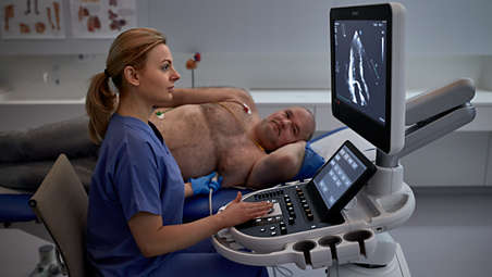
Designed to optimize cardiac workflow, the interface delivers "walk-up usability" so that users can perform exams with minimal training. The interface can be configured to match your specific workflow, which means you can focus on the patient and images, rather than searching for controls.
AutoStrain LV with automated EF and mid-layer strain

AutoStrain LV with automated EF and mid-layer strain
AutoStrain LV with automated EF and mid-layer strain
2D Auto EF and 2D Auto EF Advanced

2D Auto EF and 2D Auto EF Advanced
2D Auto EF and 2D Auto EF Advanced
Auto Segmental Wall Motion Scoring

Auto Segmental Wall Motion Scoring
Auto Segmental Wall Motion Scoring
S5-1 PureWave Transducer

S5-1 PureWave Transducer
S5-1 PureWave Transducer
X5-1 xMatrix transducer
X5-1 xMatrix transducer
X5-1 xMatrix transducer
Dynamic HeartModel

Dynamic HeartModel
Dynamic HeartModel
3D Markers

3D Markers
3D Markers
Auto Measure provides automation for robust, proven, reproducible cardiac quantification

Auto Measure provides automation for robust, proven, reproducible cardiac quantification
Auto Measure provides automation for robust, proven, reproducible cardiac quantification
iRotate to easily access an optimal view
iRotate to easily access an optimal view
iRotate to easily access an optimal view
Next-generation TEE imaging
Next-generation TEE imaging
Next-generation TEE imaging
Collaboration Live for tele-ultrasound
Collaboration Live for tele-ultrasound
Collaboration Live for tele-ultrasound
Experience echo workflow with Ultrasound Workspace

Experience echo workflow with Ultrasound Workspace
Experience echo workflow with Ultrasound Workspace
Exceptional advances for pediatric imaging
Exceptional advances for pediatric imaging
Exceptional advances for pediatric imaging
Elevated vascular assessment
Elevated vascular assessment
Elevated vascular assessment
3D Auto MV for mitral valve quantification
3D Auto MV for mitral valve quantification
3D Auto MV for mitral valve quantification
3D Auto LAA for left atrial appendage sizing
3D Auto LAA for left atrial appendage sizing
3D Auto LAA for left atrial appendage sizing
Customizable cardiology-focused interface
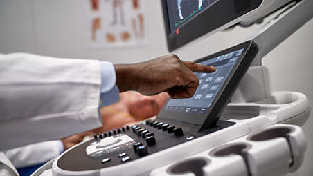
Customizable cardiology-focused interface
Customizable cardiology-focused interface
- Affiniti CVx: AutoStrain LV with automated EF and mid-layer strain
- Affiniti CVx: 2D Auto EF and 2D Auto EF Advanced (Purchasable)
- Auto Segmental Wall Motion Scoring (Purchasable)
- S5-1 PureWave Transducer
- Affiniti CVx: AutoStrain LV with automated EF and mid-layer strain
-
AutoStrain LV with automated EF and mid-layer strain
AutoStrain delivers fast, reproducible 2D strain quantification for the LV, LA, and RV, as well as automated EF and mid-layer strain for a comprehensive LV assessment within the same application, improving workflow and saving time. Smart View Select works in the background and uses AI to automatically select the optimum images for 2D LV assessment. - Affiniti CVx: 2D Auto EF and 2D Auto EF Advanced (Purchasable)
-
2D Auto EF and 2D Auto EF Advanced
Offers fast, reproducible results for easy comparison of LV functional data to improve workflow and save time in LV assessments. 2D Auto EF supports non-contrast images, and 2D Auto EF Advanced uses AI to support the quantification of contrast and non-contrast images. - Auto Segmental Wall Motion Scoring (Purchasable)
-
Auto Segmental Wall Motion Scoring
Provides automated evaluation of wall motion in a standard 17-segment bullseye display to aid objective LV wall assessment. With Auto SWMS, you can achieve greater reproducibility and efficiency in your workflows. - S5-1 PureWave Transducer
-
S5-1 PureWave Transducer
The S5-1 transducer with PureWave crystal technology generates extended operating frequency range from 5 to 1 MHz. It supports imaging in 2D, contrast mode, coronary color, CW & PW Doppler, steerable pulsed wave, High-PRF, color doppler, tissue doppler and biopsy guide capabilities. It uses reoptimized xRes adaptive image processing to provide refined tissue pattern displays, with improved details, better edges and fine border definition. It supports adult echo, abdominal, pediatric echo and TCD applications. - X5-1 xMatrix transducer (Purchasable)
-
X5-1 xMatrix transducer
Our most leading-edge, versatile ultrasound transducer technology provides remarkable 2D and 3D TTE image quality with xPlane and iRotate capabilities. Designed to simplify your imaging workflow for even difficult-to-image patients. - Dynamic HeartModel (Purchasable)
-
Dynamic HeartModel
Dynamic HeartModel brings fully automated, advanced live 3D quantification. With one button press you can get 3D EF, LV and LA quantification from the same cycle . - Affiniti CVx: 3D Markers
-
3D Markers
Graphic markers can be placed within a 3D Volume or MPR while in MultiVue to streamline tracking of structural points of interest. 3D Markers facilitate greater efficiency during echo-guided procedures and allow for detailed annotation of complex anatomy. - Affiniti CVx: Auto Measure provides automation for robust, proven, reproducible cardiac quantification
-
Auto Measure provides automation for robust, proven, reproducible cardiac quantification
Auto Measure provides fully automated 2D doppler and length measurements along with AI assisted Smart (Doppler) View ID which further enhances time-savings. AI provides full functionality without EKG. - iRotate to easily access an optimal view
-
iRotate to easily access an optimal view
Electronically access the optimum view within the acoustical window between ribs instead of manually rotating the transducer to search for an unobscured window. This can increase accuracy in measuring LV volumes because the image is less likely to be foreshortened. - Next-generation TEE imaging (Purchasable)
-
Next-generation TEE imaging
The xMatrix X8-2t transducer brings exceptional image quality and confidence to TEE imaging. Live 3D and Live 3D color flow, together with latest capabilities such as xPlane Doppler and MultiVue, help you to make a confident diagnosis in even the most complex cases. - Collaboration Live for tele-ultrasound (Purchasable)
-
Collaboration Live for tele-ultrasound
Now your ultrasound system can do more than scan. Collaboration Live allows you to reach out directly from the ultrasound system for real-time access to remote senior expertise. Collaboration Live with multi-party lets you connect up to six participants in a call. You can even connect system to system so you can give and get support from your colleagues during an ultrasound exam. - Experience echo workflow with Ultrasound Workspace
-
Experience echo workflow with Ultrasound Workspace
Ultrasound Workspace is a holistic, scalable, cardiovascular viewing, analysis and reporting system which is built on the foundation of the TOMTEC-ARENA platform. It allows for top-notch clinical efficiency by providing care teams with workflow flexibility: Enabling the same diagnostic capabilities on- and off-cart; analyzing vendor-agnostic data; leveraging AI across a wide range of applications; with a highly scalable technology platform and licensing model; and tailored comprehensive support. - Exceptional advances for pediatric imaging (Purchasable)
-
Exceptional advances for pediatric imaging
From fetal echo to pediatrics to adult congenital patients, Affiniti CVx offers a depth of imaging capability combined with streamlined cardiac workflow to reduce the steps and time needed for challenging exams such as TTE and TEE. - Elevated vascular assessment (Purchasable)
-
Elevated vascular assessment
The lightweight L12-3 ERGO transducer provides superb detail and resolution including MicroFlow Imaging for remarkable sensitivity in assessing blood flow. The eL18-4 transducer provides thin-slice imaging for exceptional tissue uniformity from near to far depth of field across a wide range of applications and depth requirements. - 3D Auto MV for mitral valve quantification (Purchasable)
-
3D Auto MV for mitral valve quantification
Analyze the complex anatomy of the mitral valve in 3D as well as its dynamic mechanics. This is useful from the initial discovery of mitral valve disease or pathology to support device planning, and through monitoring of pre- and postoperative cases. - 3D Auto LAA for left atrial appendage sizing (Purchasable)
-
3D Auto LAA for left atrial appendage sizing
Acquire the LAA ostium size quickly and easily. Automation reduces inter- or intra-user variability, increasing confidence during procedures. - Customizable cardiology-focused interface
-
Customizable cardiology-focused interface
Designed to optimize cardiac workflow, the interface delivers "walk-up usability" so that users can perform exams with minimal training. The interface can be configured to match your specific workflow, which means you can focus on the patient and images, rather than searching for controls.
AutoStrain LV with automated EF and mid-layer strain
AutoStrain LV with automated EF and mid-layer strain
AutoStrain LV with automated EF and mid-layer strain
2D Auto EF and 2D Auto EF Advanced
2D Auto EF and 2D Auto EF Advanced
2D Auto EF and 2D Auto EF Advanced
Auto Segmental Wall Motion Scoring
Auto Segmental Wall Motion Scoring
Auto Segmental Wall Motion Scoring
S5-1 PureWave Transducer
S5-1 PureWave Transducer
S5-1 PureWave Transducer
X5-1 xMatrix transducer
X5-1 xMatrix transducer
X5-1 xMatrix transducer
Dynamic HeartModel
Dynamic HeartModel
Dynamic HeartModel
3D Markers
3D Markers
3D Markers
Auto Measure provides automation for robust, proven, reproducible cardiac quantification
Auto Measure provides automation for robust, proven, reproducible cardiac quantification
Auto Measure provides automation for robust, proven, reproducible cardiac quantification
iRotate to easily access an optimal view
iRotate to easily access an optimal view
iRotate to easily access an optimal view
Next-generation TEE imaging
Next-generation TEE imaging
Next-generation TEE imaging
Collaboration Live for tele-ultrasound
Collaboration Live for tele-ultrasound
Collaboration Live for tele-ultrasound
Experience echo workflow with Ultrasound Workspace
Experience echo workflow with Ultrasound Workspace
Experience echo workflow with Ultrasound Workspace
Exceptional advances for pediatric imaging
Exceptional advances for pediatric imaging
Exceptional advances for pediatric imaging
Elevated vascular assessment
Elevated vascular assessment
Elevated vascular assessment
3D Auto MV for mitral valve quantification
3D Auto MV for mitral valve quantification
3D Auto MV for mitral valve quantification
3D Auto LAA for left atrial appendage sizing
3D Auto LAA for left atrial appendage sizing
3D Auto LAA for left atrial appendage sizing
Customizable cardiology-focused interface
Customizable cardiology-focused interface
Customizable cardiology-focused interface
Specifications
- System dimensions
-
System dimensions Width - 57.2 cm
Height - 142.2-162.6 cm
Depth - 98.3 cm
Weight - 83.6 kg
-
- Control panel
-
Control panel Monitor size - 54.6 cm
Degrees of movement - 180 degrees
Height adjustment - 20.3 cm
-
- System dimensions
-
System dimensions Width - 57.2 cm
Height - 142.2-162.6 cm
-
- Control panel
-
Control panel Monitor size - 54.6 cm
Degrees of movement - 180 degrees
-
- System dimensions
-
System dimensions Width - 57.2 cm
Height - 142.2-162.6 cm
Depth - 98.3 cm
Weight - 83.6 kg
-
- Control panel
-
Control panel Monitor size - 54.6 cm
Degrees of movement - 180 degrees
Height adjustment - 20.3 cm
-
Related products
Alternative products
-
EPIQ CVx
- AI-enabled consistency offers standardized results for both routine and specialized imaging needs
- X11-4t mini TEE fits right for more patients, in your workflow, and with a portfolio you can trust
- AutoStrain LV now features 2D automated EF and mid-layer strain
- UWS, EchoNav, and Cardiovascular Workspace: Devices and features with compatible workflow
- Collaboration Live lets you securely reach out from the ultrasound system for expert support
View product
-
EPIQ CVxi
- X11-4t along with X8-2t and VeriSight Pro allow you to make the right choice for your patient
- AI-enabled consistency offers standardized results for both routine and specialized imaging needs
- Improved efficiencies in interventional echo guidance with EchoNavigator live fusion imaging
- Efficient workflow solutions to aid a wide range of interventional procedures
View product
-
Ultrasound Workspace
- Diagnostic confidence
- Clinical efficiency
- Workflow flexibility
- Intelligent applications
- Lifetime value
View product


-
EPIQ CVx
EPIQ CVx, our premium cardiovascular ultrasound system built on our innovative, modular, industry-leading ultrasound platform, has powerful AI-based capabilities and advanced diagnostic solutions to help you transcend today's complexities and propel echocardiography into the next dimension. This enables you to achieve greater consistency, accessible innovation, smarter workflows, and easier scalability.
View product
-
EPIQ CVxi
EPIQ CVxi, our premium cardiovascular ultrasound system built on our innovative, modular, industry-leading ultrasound platform, has powerful AI-based capabilities and nSight imaging to help you transcend today's complexities and propel interventional echocardiography into the next dimension. This enables you to achieve procedural confidence with accessible innovation and smarter workflows.
View product
-
Ultrasound Workspace
Ultrasound Workspace is a holistic, scalable, cardiovascular viewing, analysis and reporting system which is built on the foundation of the TOMTEC-ARENA platform. It allows for top-notch clinical efficiency by providing care teams with workflow flexibility: enabling the same diagnostic capabilities on- and off-cart; analyzing multi-vendor data; leveraging AI across a wide range of applications; with a highly scalable technology platform and licensing model; and tailored comprehensive support.
View product
- Affiniti CVx is available in selected countries. Please consult your Philips representative for further details.
- *2020 IMV ServiceTrak™ Imaging Award winner for best ultrasound customer satisfaction, system performance and service.